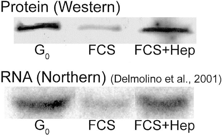
Figure 4.
Heparin maintains expression of CCN5 protein after release from G0. VSMCs were growth-arrested by serum starvation for 72 hours and subsequently reintroduced to medium containing 10% FCS/RPMI with and without heparin for 4 hours. Forty μg of each cell lysate were used for SDS-PAGE, Western blotting, and probed with anti-CCN5 peptide antibody (top). Lane 1: Lysate from G0 VSMCs. Lane 2: Lysate from cells treated with 10% FCS. Lane 3: Lysate from cells treated with 10% FCS/RPMI and 300 μg/ml of heparin. Amido Black staining (not shown) insured equal loading of protein on gel. The band representing CCN5 appears at ∼28 kd. Bottom: Northern blot analysis of CCN5 mRNA levels in VSMCs treated under exactly the same conditions used for the Western blot in the top panel. The Northern blot data are taken from Delmolino and colleagues 3 and is included here to permit direct comparison of CCN5 mRNA and protein levels.

