
Figure 3.
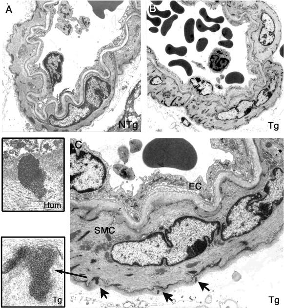
GOM deposits in cerebral blood vessels from 17- to 20-month-old transgenic mice. Ultrathin brain vessel sections from a 19-month-old nontransgenic littermate control (NTg) (A) and from a 17-month-old transgenic mouse (line Ma) (Tg) (B, C) were examined by electron microscopy. Vessel from the transgenic mouse showed no prominent changes but exhibited electron-dense granular deposits corresponding to GOM (arrows) within the basement membrane that are better seen on the higher magnification (C). Higher magnification of a GOM in a transgenic mouse brain vessel (inset, Tg) and of a GOM in a CADASIL patient brain vessel (inset, Hum) showing that GOM deposits in mouse and human had a similar structure. SMC, smooth muscle cell; EC, endothelial cell. Original magnifications: ×1293 (A, B); ×3597 (C); ×27,800 (inset, Tg); ×19,500 (inset, Hum).