
Figure 8.
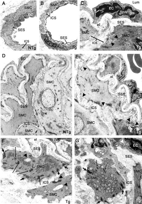
Ultrastructural changes of smooth muscle cells and of endothelial cells in tail arteries from 10- to 12 month-old transgenic mice, but no GOM deposits. Ultrathin tail artery sections of nontransgenic littermate control (NTg) (A, 10 months old; D, 12 months old) and of transgenic mice (Tg) (B and C, 10 months old; E–G, 12 months old) were examined by electron microscopy. Vessels from transgenic mice exhibited an enlarged intersmooth muscle cell space (better seen on C) and an enlarged subendothelial space filled with extracellular matrix and cellular debris (better seen on E–G). Smooth muscle cells contained more and thicker dense plaques (large arrows) having an abnormal oblique orientation and an increased number of dense bodies (thin arrows) (better seen on F and G). Starting signs of smooth muscle cell degeneration were also observed with a blurred and moth eaten appearance of the plasma membrane, the presence of some vacuoles (arrowheads) (C, E, F), or the abnormal accumulation of mitochondriae (G). Endothelial cells (ECs) with pyknotic nuclei were also detected (C, E, G). Importantly, no GOM deposit was observed. Lum, lumen; el, elastica lamina; SMC, smooth muscle cell; EC, endothelial cell; SES, subendothelial space; ICS, intersmooth muscle cell space. Original magnifications: ×1270 (A, B); ×6000 (C); ×1670 (D, E); ×4646 (G).