
Figure 2.
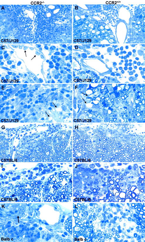
Typical acute EAE lesions in ko and wt. A–F: C57/J129 mice. Note that the inflammatory infiltrates are composed predominantly of neutrophils in CCR2−/− mice (A and C) versus monocytes in wt animals (B and D). Axonal damage is apparent as ballooned and condensed nerve fibers (WD), seen mainly in wt animals (B, D, F), and rarely in CCR2−/− (A, C, E). Arrows in C indicate high endothelial venules in ko animals. Demyelinated fibers were found in both CCR2−/− and wt animals (arrows in E and F). A: Magnification, ×250, B: Magnification, ×250, C: Magnification, ×625, D: Magnification, ×625, E: Magnification, ×625, F: Magnification, ×625. G–J: C57BL/6 mice displayed similar inflammatory infiltrates comprising neutrophils, more so in CCR2−/− (G, I), monocytes, macrophages, and lymphocytes. More severe demyelination was observed in the spinal cord of wt mice (H, J). G: Magnification, ×250, H: Magnification, ×250, I: Magnification, ×625, J: Magnification, ×625. K and L: Balb c CCR2−/− mice (K) mice displayed a preponderance of neutrophils, while wt lesions contained more monocytes (L). Note the affected root entry zone at arrow in (K). K: Magnification, ×625, L: Magnification, ×625.