
Figure 4.
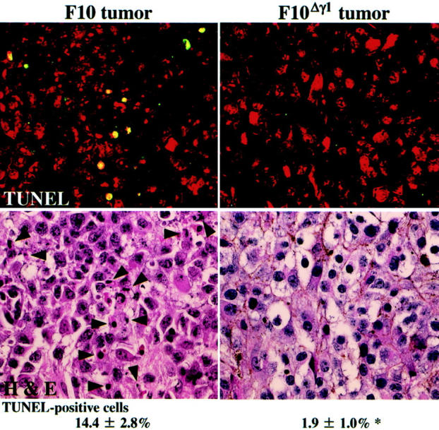
In situ detection of DNA fragmentation in footpad tumors derived from F10 and F10Δγ1 cells. Sections of the tumor tissues 4 days after irradiation were stained with the fluorescein-based TUNEL regent and PI and observed with the LSM510 microscope (top). TUNEL-positive cells contain yellow nuclei because the nuclei of all cells were also stained red with PI. After washing, the same sections were stained with H&E (bottom). Arrowheads indicate TUNEL-positive cells (top). The mean proportions of TUNEL-positive cells appear under the histological images. *, P < 0.05 by t-test when compared with the value of F10 tumors. Original magnifications, ×400.