
Figure 2.
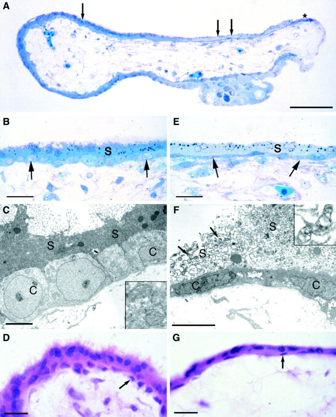
A: Light micrograph of a semithin resin section of a placental villus showing marked differences in the staining characteristics with methylene blue of the trophoblast around its circumference. The darkly staining syncytiotrophoblast on the left (single arrow) is considered healthy, whereas the pale staining area on the right (double arrow) is considered stressed. Note that the cytotrophoblast cells under the stressed syncytium are conspicuously elongated, and that they come to the surface at the extreme right-hand margin of the villus (asterisk) where they are forming a new syncytiotrophoblastic layer. B: Higher power photomicrograph of the healthy syncytiotrophoblast at the point marked by a single arrow in A. The apical border of the syncytiotrophoblast (s) carries numerous microvilli, and the syncytioplasm is uniformly dense. The underlying cytotrophoblast cells (arrows) are rounded and evenly spaced along the basement membrane. Both layers of the trophoblast show a similar staining intensity, indicating equivalent protein composition. C: Transmission electron micrograph of an area equivalent to that illustrated in B. Note the apical microvilli, the dense syncytioplasm (s), and the rounded cytotrophoblast cells (c). The syncytial mitochondria (inset) display a regular cristal arrangement. D: Light micrograph of a paraffin section of an 8 week villus demonstrating a healthy syncytiotrophoblast with numerous microvilli and an underlying complete layer of rounded cytotrophoblast cells (arrows). E: Higher power photomicrograph of the stressed area at the point in A marked by the double arrows. Note the absence of microvilli on the apical surface, the vacuolated and leached appearance of the syncytioplasm (s), and the elongated nature of the underlying cytotrophoblast cells (arrows). F: Transmission electron micrograph of an area equivalent to that illustrated in E. There is a complete absence of microvilli, and often the integrity of the apical membrane is lost. The syncytioplasm (s) is heavily vacuolated, and the underlying cytotrophoblast cells (c) are elongated along the basement membrane. The syncytial mitochondria (arrows and inset) display gross dilatation of the intracristal space, similar to that seen in severe oxidative stress in vitro. 20 G: Light micrograph of a paraffin section of an 8 week villus displaying stressed syncytiotrophoblast and flattening of the underlying cytotrophoblast cells (arrows). Scale bars: 500 μm (A); 50 μm (B, E); 5 μm (C, F); 20 μm (D, G).