Figure 4.
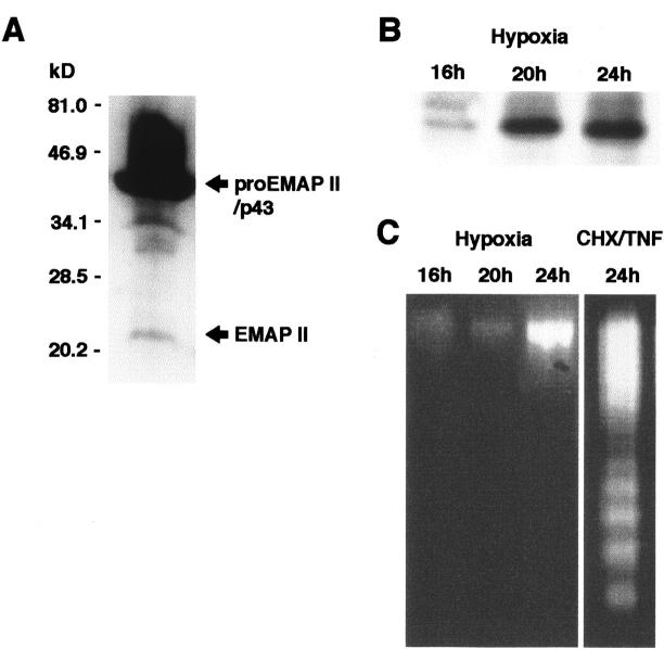
A: Expression of EMAP II protein in the B16 melanoma in vivo. Protein extracts were analyzed by Western blotting using the EMAP II-specific polyclonal antiserum SA 2847. ProEMAP II/p43 and EMAP II proteins are indicated by arrows. B: Release of EMAP II protein from hypoxic B16 melanoma cells in vitro. B16 melanoma cells were cultured at an oxygen concentration of 1% in a hypoxic chamber for the times indicated. Culture supernatants were subjected to Western blot analysis using the EMAP II-specific antiserum. Only the band corresponding to the EMAP II protein is depicted. Release of EMAP II protein under hypoxia was observed in five independent experiments. C: Hypoxic cells release EMAP II protein in the absence of DNA fragmentation. Cells corresponding to the supernatants subjected to Western blotting in B were analyzed for DNA fragmentation indicative of apoptosis (left panel). As a positive control, fragmentation of B16 melanoma cells, treated with a combination of cycloheximide and TNF (right panel) to induce apoptosis, is shown.

