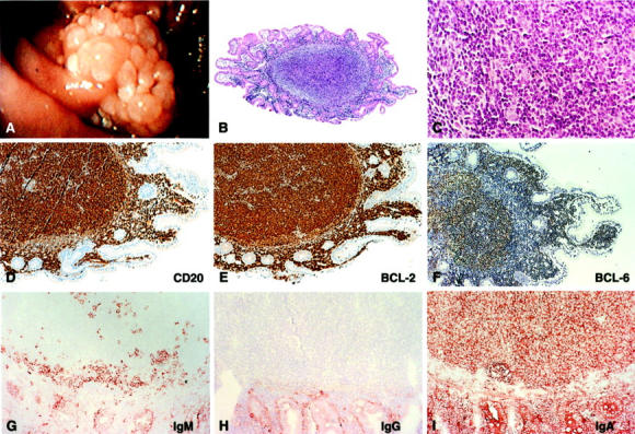
Figure 2.

Endoscopy and histology of the duodenal polyp of patient 3. A: Endoscopic picture of the adenoma-like structure found near the ampulla of Vater. B and C: H&E staining of a section through one of the tumor nodules showing a follicle-like lymphocytic infiltrate in the mucosa at ×50 and ×400 magnification, respectively. D: CD20 staining, proving the B-cell origin of the majority of the infiltrating lymphocytes. E: BCL-2 staining showing strong overexpression of this oncogene by the B cells in the follicular infiltrates. F: BCL-6 staining showing expression of this typical GC B-cell marker. G, H, and I: IgM, IgG, and IgA stainings showing that the tumor cells express IgA exclusively.
