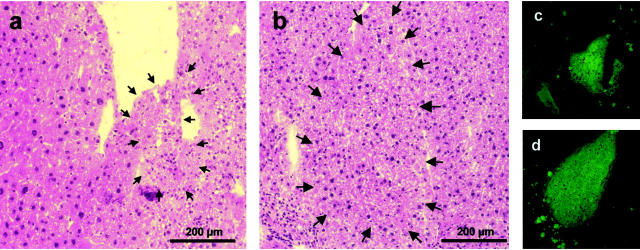
Figure 2.
Regeneration nodules after transplantation of EGFP-positive fetal liver progenitor cells in uPA/RAG-2 mice. Five-μm cryosections of uPA/RAG-2 mouse livers were stained with hemalaun-eosin and consecutive slides were analyzed for EGFP fluorescence after EGFP-positive fetal liver progenitor cell transplantation. Two weeks (a and c) after transplantation, small EGFP-positive regeneration nodules (arrows) are detectable between normal endogenously regenerated (a, left) and uPA-damaged liver tissue (a, right). After 4 weeks (b and d) the damaged liver tissue nearly was diminished and the nodules have become larger, whereby the liver morphology in the H&E staining developed a normal pattern (b). On the right part of this nodule there is a demarcation line, which is probably a result of the freezing, fixation, and staining procedure.

