
Figure 2.
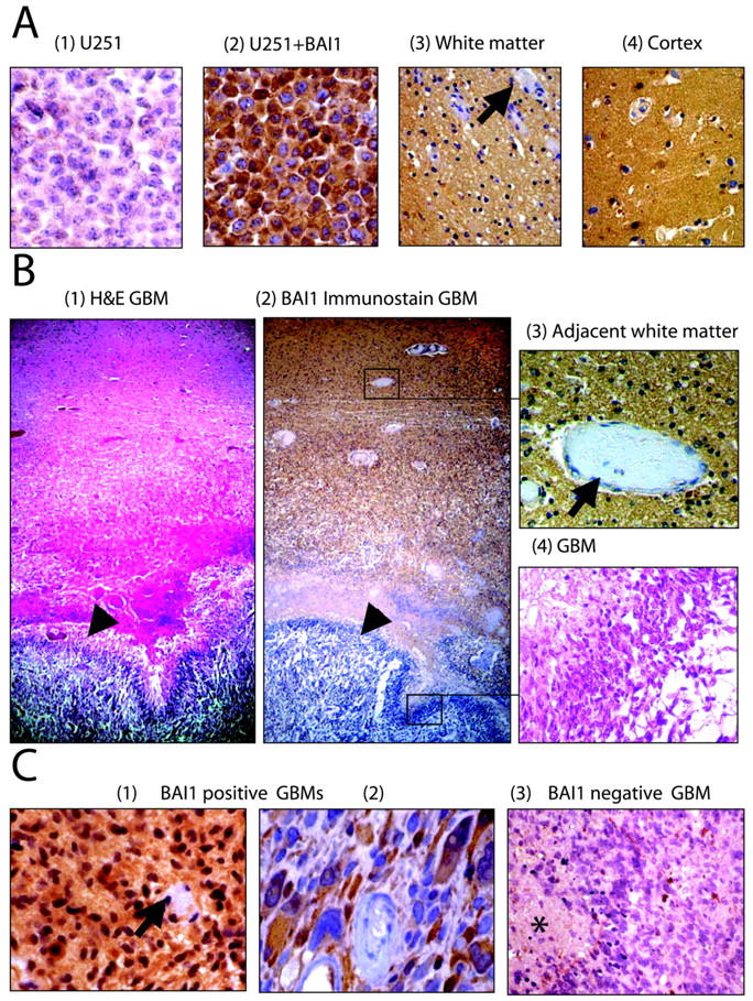
Immunohistochemistry of human glioblastoma and non-neoplastic brain specimens. A: Formalin-fixed pellets of U251MG cells untransfected (1) or stably transfected with BAI1 cDNA (2) were used as negative and positive controls for immunohistochemistry. Immunohistochemistry on non-neoplastic human brain tissue from surgically resected specimens shows diffuse expression of BAI1 in the brain parenchyma of both the white matter (3) and cortex (4). Staining is absent in vascular endothelial cells and perivascular stromal cells (arrows). B: Autopsy specimen containing GBM with adjacent non-neoplastic white matter stained by hematoxylin and eosin (1) and by immunohistochemistry for BAI1 (2). Diffuse staining of the brain parenchyma is observed in non-neoplastic brain but not in the adjacent region of GBM (arrowheads). Higher magnifications of the adjacent brain and neoplastic tissue (in boxes in 2) are shown in 3 and 4. C: Examples of GBM resection specimens showing presence (1 and 2) or absence (3) of cytoplasmic BAI1 expression in neoplastic cells by immunohistochemistry. Arrow in 1 points to vessels showing absence of expression of BAI1, and asterisk in 3 points to necrotic areas within the GBM.