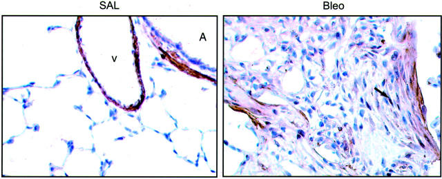
Figure 5.
Immunohistochemical analysis of ID1 expression in experimental pulmonary fibrosis. Figure ▶ shows brown immunoperoxidase staining for ID1 in insufflated normal rat lung tissue 14 days after intratracheal instillation of saline (SAL) compared with rats given bleomycin (Bleo). In the normal lung, ID1 immunoreactivity is confined to smooth muscle bundles surrounding major airways (A) and blood vessels (V) but in bleomycin-induced pulmonary fibrosis, ID1 was also immunolocalized to (myo)fibroblasts within fibrotic foci, with some cells showing nuclear staining (arrow). Original magnifications, ×400; counterstained with Mayers hematoxylin.

