
Figure 3.
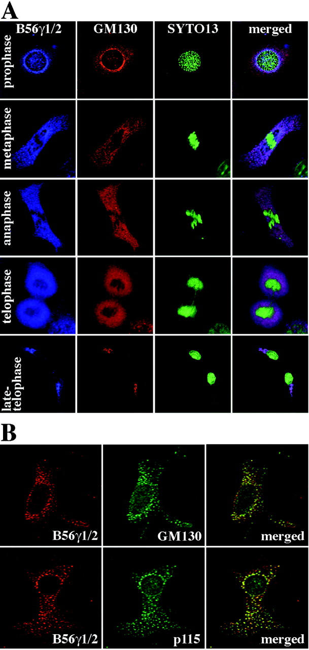
Association of endogenous B56γ subunits with cis-Golgi marker proteins during Golgi fragmentation. A: NIH3T3 cells were synchronized so that the cell population was enriched with mitotic cells. The cells were then fixed with methanol and triple-labeled with anti-B56γ1/2 Ab (Cy5, blue), anti-GM130 Ab (Cy3, red), and SYTO13 (green). The three images were then merged (right). Cells in prophase, metaphase, anaphase, telophase, and late telophase are shown. B: NIH3T3 cells were treated with nocodazole for 2 hours and double labeled with anti-B56γ1/2 Ab and either anti-GM130 Ab (top row) or anti-p115 Ab (bottom row). The anti-B56γ1/2 Ab was stained with Cy5 (red, left) and the other two Abs with Cy2 (green, middle). Cy5 and Cy2 images were then merged (right).