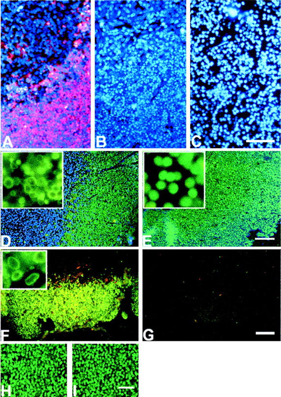
Figure 1.

A–C: Comparison of apoptotic (A, B) and necrotic (C) thymus stained by DAPI (blue fluorescence) and by in situ ligation using oligonucleotide probes with single dA overhangs (red fluorescence). A and C are dual-stained images, B is a single-stained (DAPI) image and is provided for the easy comparison of nuclear morphology with C. D and E: Comparison of apoptotic and necrotic thymus stained by TUNEL. D: Apoptotic thymus (green fluorescence, TUNEL staining; blue fluorescence, DAPI staining). E: Necrotic thymus (green fluorescence, TUNEL staining; blue fluorescence, DAPI staining). Insets show high-magnification images of apoptotic and necrotic nuclei labeled by TUNEL (side of the inset, 80 μm). Strong positive staining is present in both cases. However necrotic thymus is uniformly positive with the loss of the characteristic pattern of cell death seen in glucocorticoid-induced apoptosis. F and G: Comparison of apoptotic and necrotic thymus using in situ ligation with blunt-ended probes detecting double-strand DNA breaks bearing 5′ phosphates. F: Apoptotic thymus, blunt ends detection. G: Necrotic thymus, blunt ends detection. Note that in situ ligation does not detect double-strand DNA breaks bearing 5′ phosphates in necrotic thymus. H and I: DNA nicks relegation in necrotic thymus using T4 DNA ligase. H: Necrotic thymus TUNEL-stained before T4 ligase pretreatment; I: the same thymus after T4 ligase pretreatment. DNA nicks do not contribute to the strong positive staining of necrotic thymus by TUNEL. No change in TUNEL signal intensity occurred after treatment of necrotic tissue with T4 DNA ligase to seal DNA nicks. Apoptosis was induced in the thymus by an intraperitoneal injection of 6 mg/kg of dexamethasone. Necrosis was induced in the thymus by freezing with liquid nitrogen. Scale bars: 200 μm (C, E); 300 μm (G); 150 μm (I).
