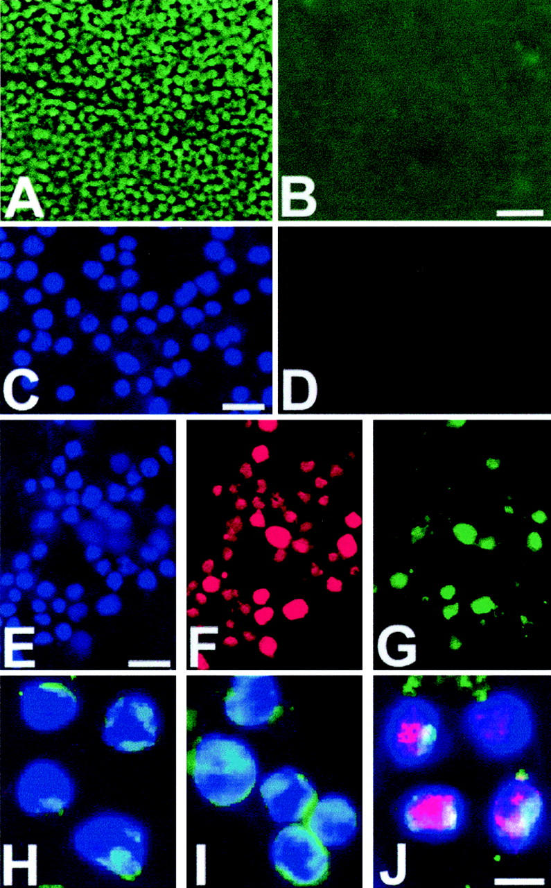
Figure 2.

Detection of 5′ overhangs but not 3′ overhangs in necrotic and apoptotic thymus and Jurkat cells in culture. A: Detection of 5′overhangs: necrotic thymus is labeled by in situ ligation (green fluorescence) with the use of a blunt-ended probe after treatment with Klenow enzyme with addition of dNTPs. B: Necrotic thymus labeled by in situ ligation with the use of a blunt-ended probe after treatment with Klenow enzyme without addition of dNTPs; no 3′overhangs detected. C: DAPI-stained necrotic Jurkat cells. D: Same cells labeled by in situ ligation with the use of a blunt-ended probe after treatment with Klenow enzyme without addition of dNTPs; no 3′overhangs detected in necrotic Jurkat cells. E, F, G: Detection of 5′overhangs. G: Necrotic Jurkat cells are labeled by in situ ligation (green fluorescence) with the use of a blunt-ended probe after treatment with Klenow enzyme with addition of dNTPs. F: Necrotic Jurkat cells are labeled by filling-in reaction using Klenow enzyme and Texas Red dUTP (red fluorescence). E: DAPI staining (blue fluorescence). H, I, J: Apoptotic thymocytes: detection of blunt ends (green fluorescence) (H), 3′overhangs (green fluorescence) (I), and 5′ overhangs (pink) (J). DAPI staining (blue fluorescence). Apoptosis was induced in Jurkat cells by the addition of staurosporine. Apoptosis was induced in the thymus by an intraperitoneal injection of 6 mg/kg dexamethasone. Necrosis was induced in the thymus by freezing with liquid nitrogen. Scale bars: 50 μm (B); 30 μm (C, E); 10 μm (J).
