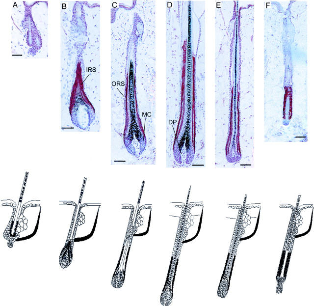
Figure 1.
PRL-like immunoreactivity during the depilation-induced murine hair cycle was stained by the ABC method using AEC+ (red) as substrate and hematoxylin for counterstaining. A, Telogen; B, anagen III hair follicles; C, anagen IV; D, anagen VI; E, catagen III; and F, catagen VII. MC, germinal matrix cells. Bottom: Schematic representation of immunoreactivity patterns of PRL during the murine hair cycle. Black: PRL expression.

