
Figure 3.
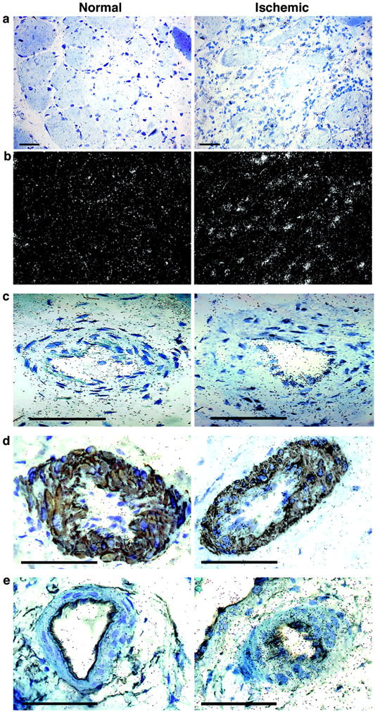
In situ hybridization analysis of ANGPTL4 mRNA levels in critical leg ischemia. Slides were counterstained with toluidine blue. The normal part of the leg is shown on the left and the ischemic part of the leg on the right. a: Bright-field view showing ANGPTL4 mRNA in tissues. b: Dark-field view showing ANGPTL4 mRNA. c: ANGPTL4 mRNA production in large vessels showing restricted labeling in the ECs and smooth muscle cells in the ischemic part. d: ANGPTL4 mRNA in SMCs in the ischemic part (right) of the leg, compared with the normal part (left), by double-labeling with an anti-smooth muscle cell actin antibody. e: ANGPTL4 mRNA in ECs of a large vessel of the ischemic part of the leg (right), compared with the normal part of the leg (left), by double-labeling with an anti-CD34 antibody. Scale bars, 100 μm.