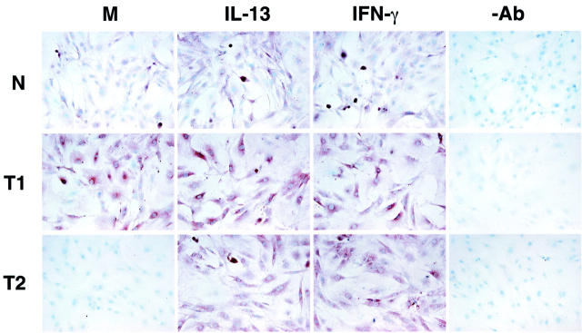
Figure 4.
Immunohistochemical analysis of IL-4Rα in tissue cultures of primary normal (N), Th1-type (T1), and Th2-type (T2) fibroblast lines. T1 fibroblasts strongly expressed IL-4Rα under basal conditions (M) compared to N and T2 fibroblasts. Cytokine treatments enhanced IL-4Rα protein expression on all three fibroblast populations. −Ab is an abbreviation representing the control antibody used.

