
Figure 5.
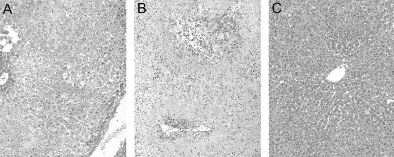
Representative histology of steatotic liver grafts harvested at day 1 after OLT. Untreated (A) and scrambled peptide-treated control (B) OLTs were characterized by extensive centrilobular pallor and necrosis with central vein vascular congestion throughout the entire biopsy. Neutrophilic infiltration was noted adjacent to the necrotic tissue. In contrast, CS1-treated OLTs (C) showed mild periportal and central ballooning changes with virtually absence of vascular congestion or necrosis (n = 3 to 4/group). H&E stain; original magnifications, ×100.