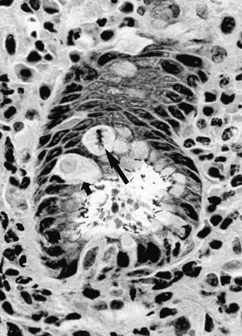
Figure 3.

Malgun cells at the proliferative zone. Note two malgun cells in the interphase (small arrow) and metaphase (large arrow), respectively. By triple silver staining, malgun cells are not stained by silver impregnation but faintly counterstained by hematoxylin. Adjacent epithelial cells are densely stained by silver impregnation. Note numerous H. pylori attached to the epithelium at the glandular lumen. Triple silver impregnation staining; original magnification, ×400.
