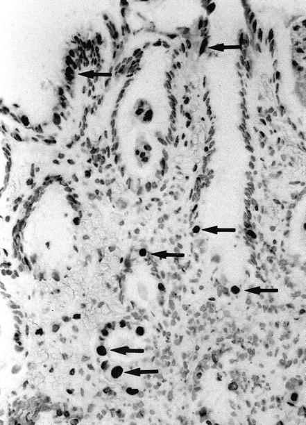
Figure 4.

TUNEL staining of malgun cells. Note numerous malgun cells stained positively in the nuclei (arrows). They have enlarged, round nuclei and abundant cytoplasm, which are typical of malgun cells. They are scattered in the proliferative and surface zone, showing a typical distribution of the malgun cell change. TUNEL staining; original magnification, ×120.
