
Figure 5.
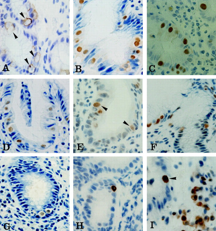
Immunohistochemical staining of malgun cells. A: Cytokeratin 8. Cytokeratin filaments are in concentric arrangement along the cytoplasmic membrane leaving a perinuclear halo of clear cytoplasm (arrowheads). Note enlarged nuclei that are only faintly stained by hematoxylin counterstaining. B–G: Ku70, PARP, proliferating cell nuclear antigen, OGG1, MSH2, and iNOS, respectively. Most malgun cells are immunostained in the nuclei. Note enlarged, round nuclei and abundant, clear cytoplasm of positively stained malgun cells. Nucleoli are seen in some malgun cells. H: p53. Note an epithelial cell strongly immunostained in the nuclei. The enlarged nuclei and prominent, unstained nucleoli are compatible with those of a malgun cell. I: bcl2. Note a malgun cell strongly immunostained in the nuclei (arrowhead). The nucleus is enlarged and the cytoplasm is abundant. Some mononuclear inflammatory cells are positively immunostained in the cytoplasm. Original magnifications: ×400 (A–C, E, I); ×240 (D, F, G, H).