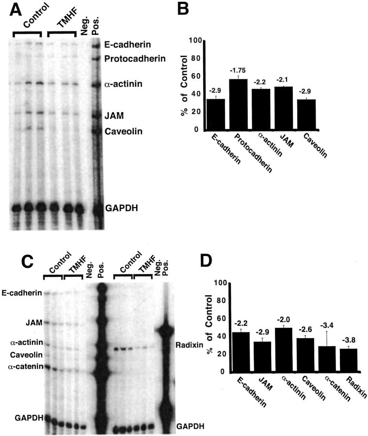
Figure 2.
RPA analysis demonstrated decreased expression of paracellular junction genes in primary rat hepatocytes after treatment with TMH-ferrocene. Primary rat hepatocytes were maintained for 7 days in control medium, after which, cultures were maintained in either control medium or medium supplemented with 20 μmol/L of TMH-ferrocene. After 21 days of treatment, control and iron-loaded cultures were harvested for total RNA. RPA analyses for the selected paracellular junction genes indicated in each figure (A and C) were performed on RNA from control (control) and iron-loaded (TMHF) cultures. Quantitative analyses of expression of selected paracellular junction genes indicated in each figure (B and D) were performed for RPA analyses for RNA from control (control) and iron-loaded (TMHF) cultures. As a control for sample processing and gel loading, the data were normalized to GAPDH expression. Each bar represents the mean percentage with SD of mRNA expression in iron-loaded cultures compared to control cultures. The fold-decrease in mRNA expression in iron-loaded cultures relative to control cultures is displayed above each bar.

