Figure 8.
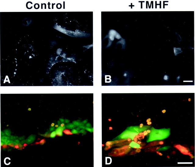
Immunolocalization of gap junction proteins and decreased GJIC in iron-loaded primary rat hepatocytes. Primary rat hepatocytes were maintained for 7 days in control medium, after which, cultures were maintained in either control medium or medium supplemented with 20 μmol/L of TMH-ferrocene for 21 days. After 21 days of iron loading, cultures were fixed and immunofluorescence was performed for connexin32 (A and B) on control (control) and iron-loaded (+TMHF) cultures. GJIC was analyzed by the scrape-loading and dye-transfer technique on control (control) and iron-loaded (+TMHF) cultures after 21 days of iron loading (C and D). Note that in control cultures (C), Lucifer yellow (green) diffused into multiple layers of cells, demonstrating functional GJIC, whereas in iron-loaded cultures (D), Lucifer yellow only diffused into a single layer of cells, demonstrating inhibition of GJIC. Original magnifications: ×400 (A, B); ×200 (C, D). Scale bars: 20 μm (A, B); 100 μm (C, D).

