
Figure 1.
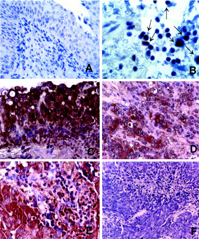
FasL expression in normal bladder urothelium and TCCs. Representative immunohistochemical stainings using the anti-FasL A11 mAb. Normal bladder: A: Normal urothelial cells are negative for FasL immunostaining. B: A higher magnification of A shows occasional stromal-positive lymphoid cells (arrows). Bladder TCC: C: FasL is expressed by all of the tumor cells that show evident membrane reinforcements. D: An infiltrating TCC, showing negative scattered tumor cells. E: Stromal FasL-positive lymphoid cells in a FasL-positive tumor. F: A FasL-negative high-grade TCC. Immunostaining is in brown. Counterstain with hematoxylin. Original magnifications: ×20 (A, F); ×40 (C, D, E); ×100 (B).