
Figure 6.
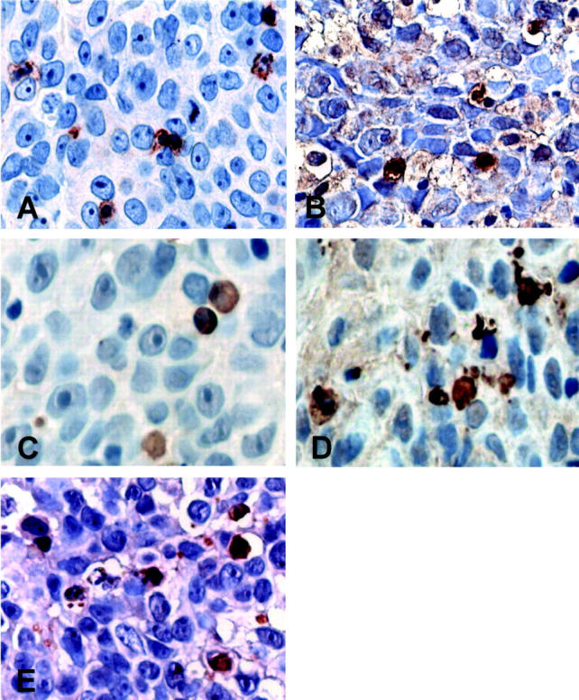
Caspase-8, -9, and -3 expression by IFN-γ-producing CD8-positive TILs in a FasL-positive TCC. Larger magnifications of central tumor areas depicted in Figure 5 ▶ are shown. A: Surface CD8-positive cell staining appeared clump-like. CD8-positive cells had large cytoplasms and coarctate nuclei; B: Some of the CD8-positive T lymphocytes, located in close contact with the tumor cells, were strongly positive for IFN-γ immunostaining. Caspase-8 (C)-, caspase-9 (D)-, and caspase-3 (E)-positive cells showed a tissue distribution and morphology comparable to that of IFN-γ- and CD8-positive T lymphocytes. Immunostaining is in brown. Counterstain with hematoxylin. Original magnifications, ×800 (A–E).