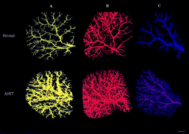
Figure 2.
The brightest voxel projection of the intrahepatic biliary tree (A), hepatic artery (B), and portal vein (C) from left lateral lobe of normal (top) and ANIT-fed (bottom) rats reconstructed in three dimensions. Images are viewed at 80° increments, but any other axial direction could be selected. The images clearly demonstrate significant bile duct proliferation and hepatic artery and portal vein neovascularization after ANIT feeding. All structures developed well-organized networks connected with pre-existing networks. Scale bar, 2 mm.

