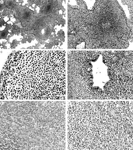
Figure 3.

Pulmonary MALT lymphomas showing “typical” (A–D) or “atypical” (E and F) histology. A: The neoplastic lymphocytic infiltrate extends into the pulmonary parenchyma along bronchovascular septa. B: A lymphoid follicle with intact mantle zone is surrounded by a diffuse small lymphoid infiltrate. C: Tumor cells show indented or folded nuclei and a moderate amount of pale cytoplasm. D: Tumor cells infiltrate in the bronchial mucosa forming a lymphoepithelial lesion. E: A pulmonary MALT lymphoma with “atypical” histology showing marked plasmacytic differentiation. A non-neoplastic lymphoid follicle is seen in the left lower corner. F: A MALT lymphoma with “atypical” histology showing an increased number of large cells involving the bronchial glands (right). H&E: original magnifications; ×8 (A), ×33 (B), ×50 (C), ×100 (D), ×100 (E), ×66 (F).
