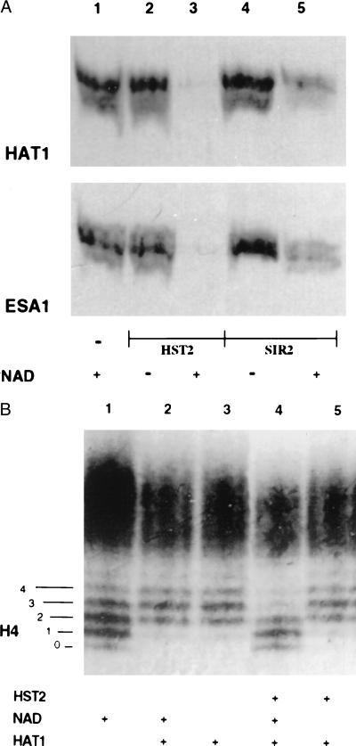
Figure 3.
Histone deacetylation analyzed by gel electrophoresis. (A) An SDS/PAGE gel of histones acetylated with [3H]acetyl-CoA by HAT1 or by ESA1, as indicated, and then treated with no enzyme (lane 1), with HST2 (lanes 2 and 3), or with SIR2 (lanes 4 and 5). A fluorogram of the gel is depicted. (B) A Triton–acid–urea gel of chicken erythrocyte histones acetylated with HAT1 followed by deacetylation by HST2. The gel was stained with Coomassie blue to visualize the histone isoforms. The numbers 0–4 on the left refer to isoforms of H4 differing in charge, with 0 corresponding to the most positively charged isoform and 4 corresponding to the least positively charged form. Lane 1, histones incubated without HAT1; lanes 2 and 3, histones acetylated with HAT1, in the presence or absence of NAD; lanes 4 and 5, histones acetylated by HAT1, incubated with HST2, with and without NAD.

