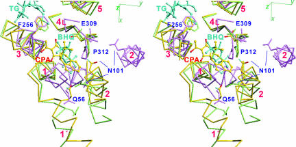
Fig. 4.
Three transmembrane inhibitors (CPA, TG, and BHQ) superimposed with Cα traces of the three crystal structures, E1·2Ca2+ (violet), E2(CPA+CC) (yellow), and E2(TG+BHQ) (light green), viewed in stereo. The side chains of several key residues are shown. The transmembrane helices (M1–M6) are numbered.

