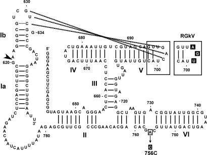
Fig. 5.
Secondary structure of RG and mutant derivatives. Nucleotides and helices are numbered as in ref. 14. Nucleotides involved in the peripheral loop–loop kissing interaction are connected with dotted lines. The cleavage site is indicated with an arrowhead. Regions that have been mutated in other constructs are enclosed in boxes, and the mutation(s) is highlighted in black (see Materials and Methods).

