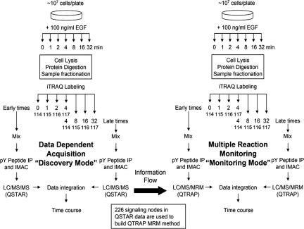
Fig. 1.
Schematic of the combined IDA/MRM method. Parental HMECs were treated with 100 ng/ml EGF for 0, 1, 2, 4, 8, 16, or 32 min after 12-h serum starvation. Samples were divided up into “early” or “late” halves of the time course, with the 4-min sample being present in both subsets as a normalization point. Multiple IDA analyses on the QSTAR mass spectrometer were used to identify nodes within the network and to obtain information needed to construct the MRM methods. After the MRM methods were constructed, identical samples from two biological replicates were analyzed by both IDA on the QSTAR and MRM on the QTRAP to compare results and estimate the quality of the MRM/QTRAP data set.

