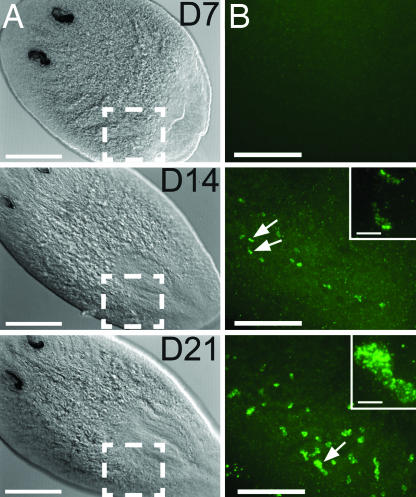
Fig. 2.
Smed-nanos expression in regenerating head fragments amputated anterior to the ovaries. (A) Differential interference contrast microscopic images of regenerating heads fixed 7, 14, or 21 days after amputation (animals were ≥1.2 cm when amputated). The numbers of animals in which nanos mRNA was detected were 7 of 14 at 7 days, 8 of 8 at 14 days, and 8 of 9 at 21 days. (Scale bars, 250 μm.) (B) Confocal projections corresponding to the boxed regions in A showing nanos mRNA detected by FISH. (Scale bars, 100 μm.) Arrows indicate nanos-positive cells shown at higher magnification in the Insets. (Inset scale bars, 10 μm.)

