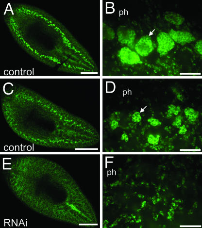
Fig. 5.
Smed-nanos RNAi knockdown animals do not regenerate testes primordia. Animals were fixed 14 days after amputation posterior to the ovaries and processed to detect germinal H4 mRNA by FISH. (A–D) Control animals. (E and F) nanos RNAi animals. (B, D, and F) Confocal images corresponding to the postpharyngeal regions of the planarians shown in A, C, and E. Control animals regenerated germinal H4-positive testes primordia (n = 10). (A and B) Well developed testes lobes (arrow in B) were observed in the largest of these specimens. (C and D) The remaining animals developed smaller clusters of germinal H4-positive testes primordia (arrow in D). (E and F) germinal H4-positive dorsal clusters were not detected in nanos RNAi animals (n = 11); only somatic neoblasts were observed. ph, pharynx. [Scale bars, 500 μm (A, C, and E); 50 μm (B, D, and F).]

