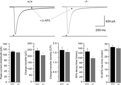
Fig. 2.
Rapid NMDA currents evoked by glutamate photolysis are normal in CA1 pyramidal cells derived from cPLA2α−/− mice. Hippocampal slices were bathed in Mg2+-free aCSF supplemented with MNI-glutamate and antagonists of GABAA, AMPA, kainate, and mGluR1/5 receptors. In these conditions, 10-msec-long flashes of UV light produced glutamate photolysis, which evoked an NMDA receptor-mediated inward current. The current traces shown are the averages of five consecutive responses. The lower trace illustrates the duration of the UV flash. These responses were completely blocked by the NMDA receptor antagonist D-AP5. Bar graphs show population comparisons. No significant differences were observed between +/+ and −/− groups (P > 0.10 for all). n = 29 and 32 cells, respectively.

