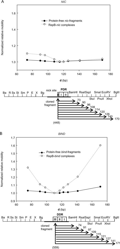
Figure 5.
RepB-induced DNA bending characterized by circular permutation assays. Both 241-bp nic- (A) and 243-bp bind- (B) permuted fragments were generated by restriction with the enzymes indicated in the scheme. Below, the distance (d, in bp) from the centre of the cloned pMV158 DNA (coordinate in brackets) to the right end of each fragment is displayed. The electrophoretic mobility of free (closed circles) or bound (open circles) DNA fragments, normalized to the mobility of the slowest same-sized DNA or complex, was plotted versus d. The apparent angles of both the intrinsic curvature and the RepB-induced bending on the nic and bind loci are shown in Table 1.

