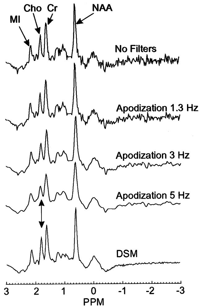
FIG. 4.
Representative 1H MR spectrum from parietal cortical gray matter from a 74-year-old CN subject. The spectrum is shown unfiltered (top), after apodization filtering using different widths (middle), and after DSM filtering (bottom). Heavy apodization filtering degraded spectral resolution, in contrast to DSM (see arrow).

