Figure 2.
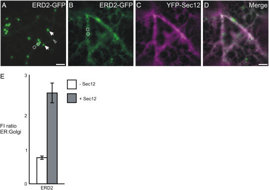
Overexpression of Sec12-YFP inhibits transport of ERD2-GFP to the Golgi apparatus. A, Confocal image of a cell expressing ERD2-GFP, which labels the Golgi apparatus (arrows) and ER (arrowhead). B to D, On coexpression of Sec12-YFP (C), ERD2-GFP is partially redistributed to the ER (B and D). Bars = 5 μm; circles represent the size of the areas used to measure fluorescence intensity in the ER and Golgi. E, Quantification of the ER-localized ERD2-GFP fluorescence relative to that in the Golgi apparatus in the absence (−Sec12) or presence (+Sec12) of Sec12-YFP. A significant increase in the ratio was verified when Sec12-YFP was expressed in comparison with the control. Sample size: 240 Golgi bodies and 240 ER measurements were analyzed for each sample. Error bars represent se of the mean.

