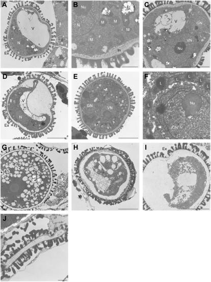
Figure 6.
Transmission electron microscopy analysis of anthers in the SDH1-1/sdh1-1 mutant. Ex, Exine layer; GN, generative nucleus; L, lipid body; M, mitochondrion; N, nucleus; Nu, nucleolus; S, starch granules within plastids; V, vacuole; VN, vegetative nucleus. Bar = 5 μm in A, C to E, and G to I; bar = 1 μm in B, F, and J. A, Microspores at the vacuolated stage (anther stage 9 according to Sanders et al., 1999). All microspores had wild-type morphology, with numerous mitochondria, starch granules within plastids, and a normal exine outer layer. B, Closeup of the microspore shown in A. C, Bicellular pollen grain from SDH1-1/sdh1-1 anthers. The asymmetric first mitotic division has occurred (early anther stage 11). D, Same anther as in C. In addition to pollen with wild-type morphology (C), abnormal pollen grains were present. They did not undergo mitosis and cell content detached from the outer layers. E and F, Later developmental stage of wild-type pollen grains from SDH1-1/sdh1-1 anthers. G, Mature wild-type pollen grain from SDH1-1/sdh1-1 anthers. H and I, Later developmental stages of aberrant pollen grains from SDH1-1/sdh1-1 anthers. Cellular structures and content progressively disappeared and internal structures were no longer distinguishable. J, Collapsed pollen grain in the same anther as in G. The exine layer, of sporophytic origin, remained wild-type like.

