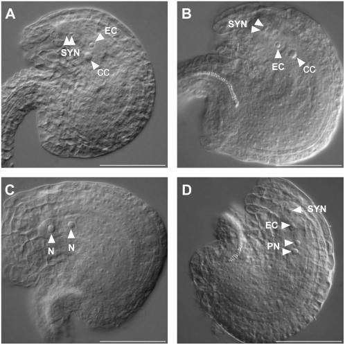
Figure 7.
Embryo sac development defects in SDH1-1/sdh1-1 mutant plants. Flowers of wild-type and SDH1-1/sdh1-1 plants were emasculated, and whole-mount preparations of ovules were analyzed by DIC microscopy 48 h after emasculation. Embryo sac nuclei are indicated by arrows. A, Ovule from a wild-type Ws plant. The embryo sac is at the terminal developmental stage and contains one secondary nucleus in the central cell, one egg cell nucleus, and two synergid cell nuclei at the micropylar end. B, Ovule harboring a wild-type-like embryo sac in a SDH1-1/sdh1-1 mutant plant. C, Ovule from the same pistil of the ovule shown in B. The embryo sac displays a mutant phenotype, being arrested at the two-nucleate stage. D, Ovule from the same pistil of the ovule shown in B and C, but showing a second mutant phenotype. The two polar nuclei lie side by side but failed to fuse. CC, Central cell nucleus; EC, egg cell nucleus; N, nuclei from an embryo sac arrested at the two-nucleate stage; PN, polar nuclei; SYN, synergid cell nuclei. Bars = 50 μm.

