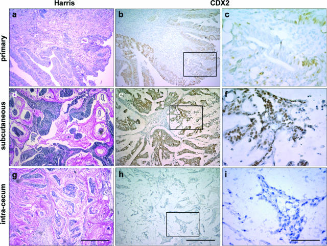
Figure 1.
CDX2 expression in human colorectal tumors. a–c: An example of human colorectal tumor at the time of surgical resection is illustrated, showing heterogeneous CDX2 expression. d–i: The corresponding tumor cells were implanted subcutaneously (d–f) or in the cecum wall (g–i). Sections were stained with Harris solution for histopathological examination (a, d, g) or immunostained with anti-CDX2 antibody (b, c, e, f, h, i). The regions enclosed in b, e, and h are shown at higher magnification in c, f, and i, respectively. Scale bars = 200 μm (a, b, d, e, g, h); 40 μm (c, f, i).

