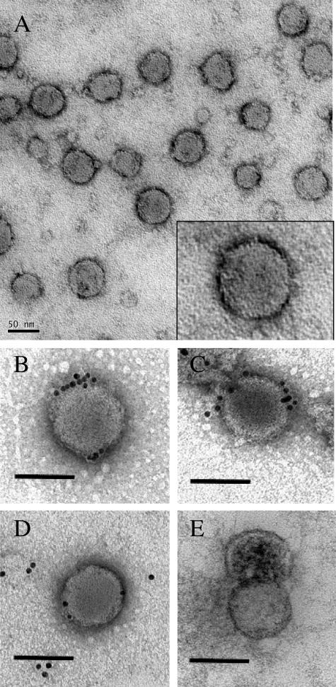
Figure 7.
Electron microscopy of virus-like particles from media of infected cultures. A sample of medium, filtered through a 1-μm membrane, containing approximately 106 HCV copies/ml was deposited by ultracentrifugation on the grids and negatively stained with 0.1% uranyl acetate (A and high magnification inset). The medium was obtained from the infected cultures shown in Figure 5B 5 days after infection. For immunogold staining, the grids were incubated with goat polyclonal antibody against HCV genotype 1a E2 (B and C), goat antibody against mouse IgG (D), or no primary antibody (E). Rabbit anti-goat IgG conjugated with 10-nm gold particles was used as the secondary antibody for all of the samples. Note the distribution of gold particles decorating a viral particle in B and C, scattered gold particles in D, and no gold labeling in E. In E, the nucleocapsid of a viral particle surrounded by an envelope is stained by uranyl acetate, probably as a consequence of the high-pressure ultracentrifugation used to deposit the particles into the grid. Scale bars: 50 nm (A); 100 nm (B–E).

