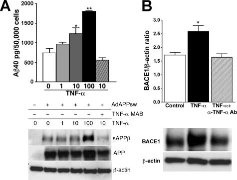
Figure 6.
TNF-α-stimulated astrocytes enhance Aβ production and BACE1 expression. A: Top: Aβ40 ELISA of tissue culture media collected from astrocyte primary cultures isolated from WT or GRKO neonates 24 hours after APPsw adenovirus infection and TNF-α (1, 10, and 100 ng/ml) stimulation or APPsw adenovirus infection and 10 ng/ml TNF-α plus 10 μg/ml α-TNF-α mAb stimulation. Bottom: The same set of tissue culture media (100 μl) was subjected to immunoprecipitation with 20 μg of 6E10 mAb (against Aβ1-17 sequence) overnight at 4°C to deplete Aβ and sAPPα. Supernatant after the immunodepletion was subjected to immunoblotting using anti-HA antibody, showing sAPPβ. Expression of full-length APP (APP) was examined by cell lysates. Expression of β-actin was used as a loading control. B: Cell lysates from astrocyte primary cultures isolated from WT or GRKO neonates stimulated by murine TNF-α (10 ng/ml) with or without anti-TNF-α antibody (α-TNF-α mAb, 10 μg/ml) for 24 hours, were subjected to immunoprecipitation with anti-BACE1 polyclonal antibody. The immunoprecipitated fraction was subjected to immunoblotting using anti-BACE1. The BACE1 band intensity was normalized against the β-actin level in the original cell lysate.

