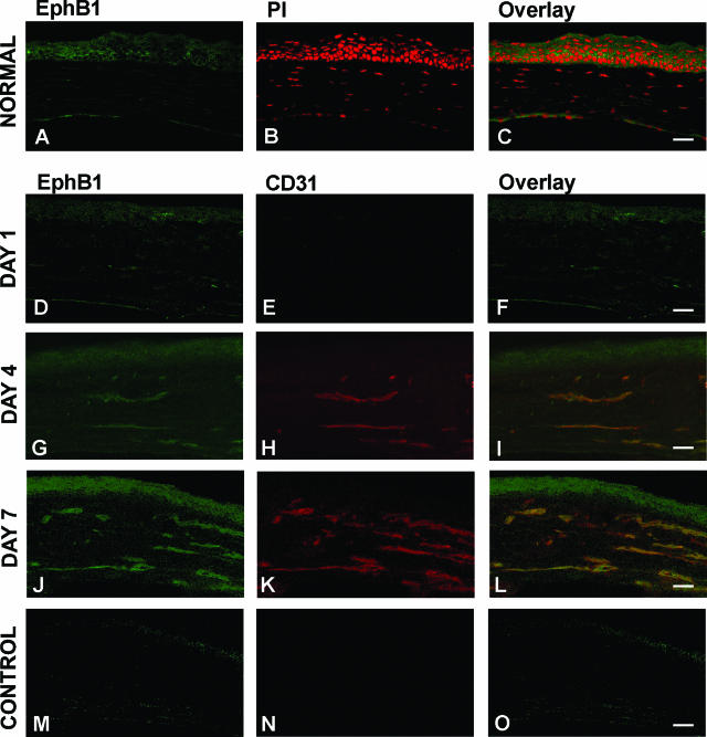
Figure 3.
EphB1 immunolocalization in normal and bFGF-induced neovascularized cornea. Uninjured normal eye was enucleated and stained with EphB1 (A) and propidium iodide (B). Overlay image was shown in C. After bFGF pellet (120 ng/pellet) insertion, mouse eyes were enucleated at days 1, 4, and 7 and stained with EphB1 (green) (D, G, and J) and CD31 (red) (E, H, and K) antibodies. Overlay images are shown in F, I, and L. Bar = 20 μm. Antibodies preincubated with cognate peptides were used as control (M–O).

