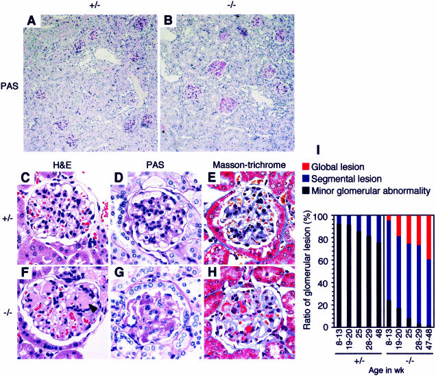
Figure 2.
Expansion of the mesangial matrix and deposition of immune complexes in the mesangial area in β4GalT-I−/− mice. A and B: Low-magnification views of PAS-stained renal cortex. C and F: H&E staining. D and G: PAS staining. E and H: Masson-trichrome staining. A, C, D, and E: β4GalT-I+/− mice at 16 months of age. B, F, G, and H: β4GalT-I−/− mice at 16 months of age. Acellular eosinophilic lesions are shown by an arrowhead in F. I: The degrees of glomerular lesion are shown at indicated ages. The segmental lesion includes segmental glomerular sclerosis, focal adhesion, and small crescent formation, whereas the global lesion includes global mesangial matrix expansion.

