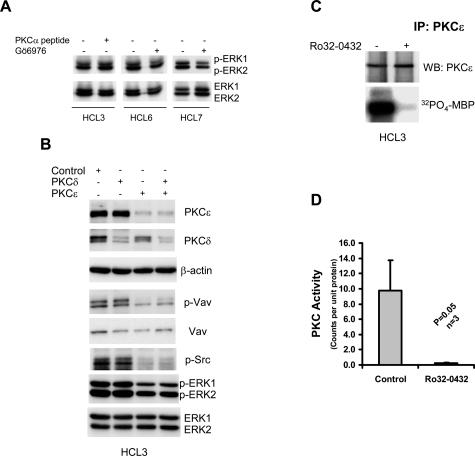
Figure 4.
The constitutive activation of ERK in HCs involves PKCε. A: Cell lysates were analyzed for the presence of phosphorylated ERK in HCs treated with 10 μmol/L PKCα-blocking peptide or with 100 nmol/L Gö6976 for 1 hour. This figure shows the results of one of three independent experiments using the PKCα-blocking peptide and two of five independent experiments using Gö6976. B: Lysates from HCs treated with control siRNA or with siRNA specific for PKCε and PKCδ were analyzed for the presence of phosphorylated Src, Vav, and ERK. Data in this figure represent one of three independent experiments with similar results. C: Immune complex kinase assay of PKCε immunoprecipitated from HC lysates performed in the presence or absence of Ro32-0432. D: A summary of the results presented in C, representing the data generated using three different HC cases. Statistical analysis was performed using a Student’s t-test.

