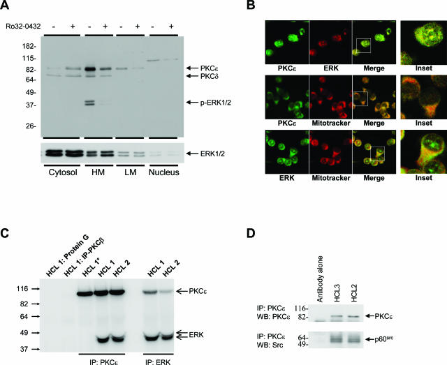
Figure 5.
PKCε co-localizes with ERK and p60src. A: HCs were fractionated into cytosolic, light membrane, heavy membrane (containing the mitochondria), and nuclear cell compartments by differential centrifugation. Equal protein amounts (10 μg) were separated by SDS-PAGE, and Western blots were probed with the indicated antibodies. Data in this figure represent one of three independent experiments with similar results. B: Confocal microscopy showing co-localization of PKCε and ERK and their association with mitochondria in HCs. Insets are magnifications of representative cells in the merged images. These images are representative of the malignant cells from two cases of HCL. C: Lysates of cells from two cases of HCL were immunoprecipitated with the indicated antibodies or with protein G-Sepharose and analyzed by Western blot for the presence of PKCε and ERK. The immune complexes in lane HCL 1* were washed with RIPA buffer before gel loading to disrupt the molecular association between PKCε and ERK. D: PKCε was isolated from cell lysates from two HC cases by immunoprecipitation and analyzed by Western blot for PKCε and p60src. C and D are representative of at least two experiments using cells from different HCL cases.

