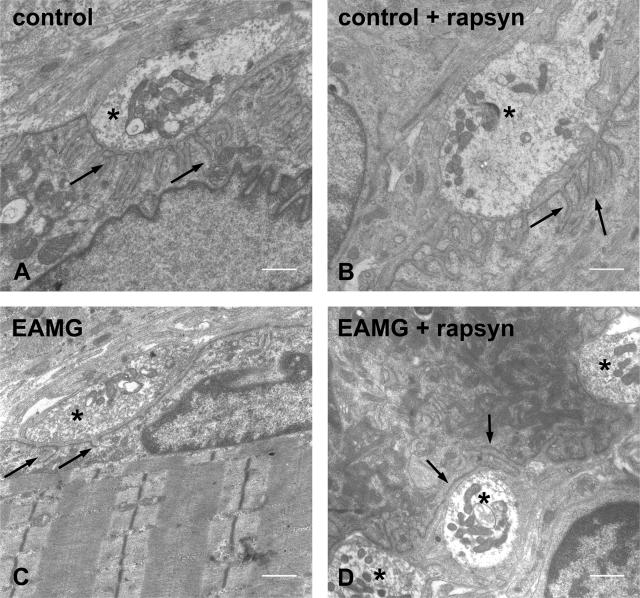
Figure 7.
Electron microscopic examinations of the postsynaptic folds of motor endplates. NMJ in an untreated (A) and rapsyn-treated (B) tibialis anterior muscle. Damaged postsynaptic membrane without folds in an untreated tibialis anterior muscle (C) and in a rapsyn-treated tibialis anterior muscle (D) of an EAMG animal. Arrows indicate the postsynaptic membranes; asterisks indicate the nerve terminals. Scale bars = 1 μm.

