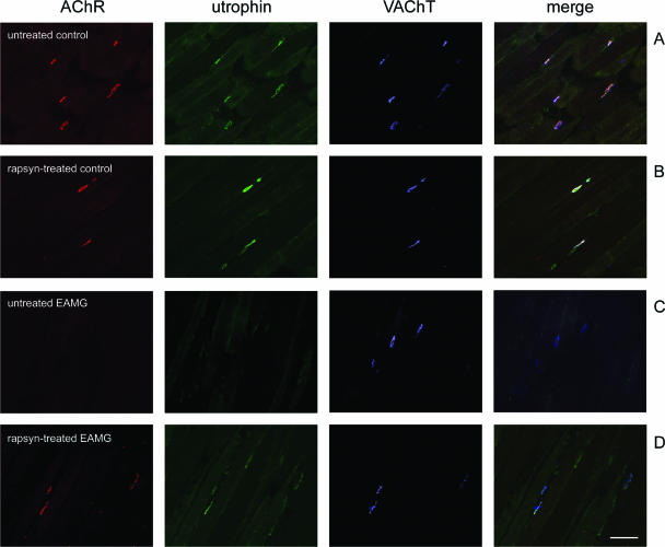
Figure 8.
Cryosections of tibialis anterior muscles were stained with α-BT (red), mouse anti-utrophin mAb MANCHO 7 (green), and rabbit anti-VAChT (blue); merge on the right. A and B: In untreated and rapsyn-treated tibialis anterior muscles of a control rat, utrophin and VAChT are co-localized at the endplates. C: In chronic EAMG muscles, endplates showed reduced staining of utrophin but not VAChT. D: In rapsyn-treated EAMG muscles, the endplates with increased amount of AChR also stained intensively for utrophin. Scale bar = 100 μm.

