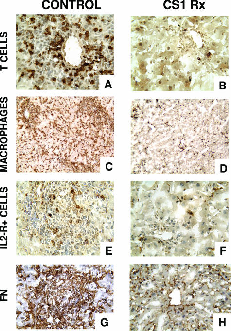
Figure 4.
Mononuclear cell infiltration/activation and fibronectin deposition in steatotic OLTs. Immunoperoxidase staining of T lymphocytes (A, B), monocyte/macrophages (C, D), IL-2R+ cells (E, F), and cellular FN (G, H) in fatty liver grafts at day 7 post-OLT. Blockade of the α4β1-FN interactions (B, D, F, H) was associated with significant decrease in intragraft infiltration of T lymphocytes, monocytes/macrophages, and IL-2R+ cells and lower levels of FN expression compared with respective control OLTs (A, C, E, G), which were characterized by massive leukocyte infiltration/activation and FN deposition. Original magnification: ×200 (A, B, E–H); ×100 (C, D) (n = 5/group).

