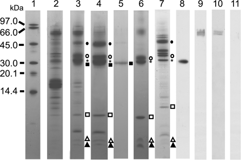
Fig. 2.
SDS/PAGE and Western blot analysis of PSII complexes isolated from A. marina. Lane 1, molecular marker; lane 2, thylakoid membranes; lane 3, partially purified PSII preparations after DEAE Toyopearl chromatography; lane 4, purified PSII complexes; lane 5, CP43′; lane 6, PSII RC isolated from spinach; lane 7, PSII core complex isolated from Synechocystis; lane 8, Western blot of purified PSII complex (lane 4) with anti-D1; lanes 9–11, Western blot with anti-PsaA/B; lane 9, thylakoid membranes (lane 2); lane 10, partially purified PSII (lane 3); lane 11, purified PSII complex (lane 4). Individual marks represent CP47 (filled circles), D2 (open circles), D1 (asterisks), CP43′ (filled squares), cyt b559 α-subunit (open squares), PsbI (open triangles), and cyt b559 β-subunit (filled triangles).

